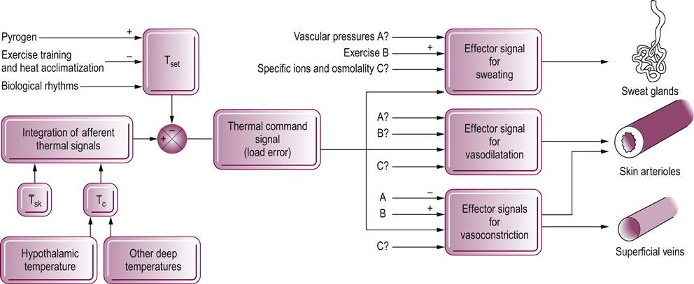
 Thermoregulation: considerations for aging people | Musculoskeletal Key
Open Access is an initiative that aims to do free scientific research for all. To date our community has made more than 100 million downloads. It is based on principles of collaboration, unhindered discovery and, most importantly, scientific progression. As PhD students, we found it difficult to access the research we needed, so we decided to create a new Open Access editor that level the playing field for scientists around the world. How? By facilitating research, it puts the academic needs of researchers to the business interests of publishers. We are a community of over 103,000 authors and editors from 3,291 institutions covering 160 countries, including Nobel Prize winners and some of the world's most recited researchers. Publishing on IntechOpen allows authors to win appointments and find new collaborators, which means that more people see their work not only from their own field of study, but also from other related fields. Brief introduction to this section that you decibes Open Access especially from an IntechOpen perspectiveDo you want to get in touch? Contact our main office in London or Our team is growing all the time, so we are always looking for smart people who want to help us reshape the world of scientific publication. Revised open access chapter by pairs Anatomy and function of the hypothalamus By Miana Gabriela Pop, Carmen Crivii and Iulian OpincariuSubmitted: June 6th 2017Reviewed: August 6th 2018Published: November 5th 2018DOI: 10.5772/intechopen.80728 √≥title Downloaded: 2683Abstract Hypothalamo is a small but important brain area. Through its neuronal connections, it is involved in many complex functions of the organism such as the control of the vegetative system, the homeostasis of the organism, thermoregulation, and also in the adjustment of emotional behavior. Hypothalamus is involved in different daily activities such as eating or drinking, in the control of the temperature and energy maintenance of the body, and in the process of memorization. It also modulates the endocrine system through its connections to the pituitary gland. The exact anatomical description along with a correct characterization of the components structures is essential to understand their functions. Palabras claves y autor infoAutoresMiana Gabriela Pop*Carmen CriviiIulian Opincariu*Add all correspondence to: miana_my@yahoo.comDOI: 10.5772/intechopen.80728 From the edited volume Edited by Stavros J. Baloyannis and Jan Oxholm Gordeladze1. Intoxication development of hypothalamus At the end of the fourth week of embryonic development, the neural tube is organized in the primary vesicles: the antebrain or prosencephalon vesicle, the mid-brain or mesencephalon vesicle, and the spinal cord, also called rombencephalon. Prosencephalon is divided into two secondary vesicles, the telencephalon that will form the cerebral hemispheres and the diencephalon that gives rise to the diencephalon. Mesencephalon forms the middle brain, structure involved in vision and hearing processes. The Hindubrain gallbladder or rombencephalon is divided into metencephalon, which forms more the ponies and the cerebel and the honeyencephalon that forms the medulla. The embryological concepts related to the development of the hypothalamic region are over 100 years old. Since Herrick [] first proposed the cylindrical model of the antebrain organization, the anatomical description was accepted per se and very few research documents have questioned its validity. The cylindrical morphological model is based on the division of the antebrain into functional longitudinal units, placing the telencephalon in the most rostral region and the diencephalon flowly, between the telencephalon and the midbrain, while the hypothalamus if formed from the ventral most of the diencephalic vesicle []. In recent decades, the mapping of the genes involved in hypothalamic development has allowed us to identify a disparity between the morphological and classic boundaries of this region and the molecular ones. According to the Prosomeric model of Puelles [], the longitudinal axis initially proposed from the brain is bent due to the first mesencephalic bending of the embryo. This condition puts the rostrally diencefallon between the cranial telencephalon and the midbrain flowly and establishes the independent hypothalamus of the diencephalon as a later part distinct from the forebrain [, ].An important role in hypothalmic development is also assigned to the presence of specific signaling centers (proliferation of morphogenic int proteins, hedgeogenic Hypothalamus is a small central region of the human brain formed by nerve fibers and a conglomerate of nuclear bodies with various functions. Hypothalamus is considered a link between the nervous system and the endocrine, its main function being to maintain the homeostasis of the body. The hypothalamus is located under the talamo of which is separated by the hypothalamus sulcus of Monro. The sulcus is on the side wall of the third ventricle and extends anteroposteriorally from the Monro interventricular foramen (which ensures the communication between the third, diencephalic ventricle and the front horn of each lateral ventricle) to the Sylvius cerebral aqueduct level. The hypothalamus is previously limited by the lamina terminalis, a layer of triangular grey matter extended above the optional chiasma, between the two previous horns of the fornix. Lamina terminalis also forms the previous wall of the third ventricle and contains the organum vasculosum, a circumventricular structure characterized by the absence of the blood-brain barrier and therefore highly sensitive to osmotic variations of the blood []. The upper wall of the hypothalamic region participates in the formation of the inferolateral wall of the third ventricle of the brain and has close relations with the structure of white matter that surrounds it, called the Fornix. The fornix is a C-shaped white brain structure that connects several parts of the brain (hypotamic nucleos with hypocampal region, talalic nuclei with mammal bodies of hypothalamus). Even if your function is not clearly understood, your relationship with memory is known, and recent studies are testing your deep brain stimulation as a treatment in advanced Alzheimer's disease []. Subsequently, hypothalamus extends to the periaqueductal grey substance and tegmentum of the upper part of the brain trunk. Only on the lower surface of the brain, hypothalamus can be visualized from the optical chiasm and the perforated substance prior to the later cerebral peduncles of the middle brain and mammal bodies, dorsally (). Mammary bodies are small and round white matter structures that belong to the lymbic system. They are involved in memory due to their connections to the hypocampal region and also in maintaining the direction sense []. The hypothalamus is laterally limited by optical pathways in its direction to the lateral geniculate bodies, an important relay of the optical path. Within the area delimited on the outer surface of the brain, a small prominence, called tuber cinereum or infundibulum connects the hypothalamus with the posterior lobe of the lower pituitary gland. Hypophysis or hypophysis is found at the base of the brain, in a depression of the sphenoid bone called a turcical seal. Figure 1. Lower surface of the brain with hypotalamic visualization at this level. 2.1. Hypothalamus: Hypophysial Complex The pituitary gland is a three-lobed structure: anterior, posterior and intermediate lobe, with different embryonic origin. The previous lobe, previous pairs, or adenohypophysics is derived from the previous wall of the Rathke bag, an ectodermal structure that also forms the primitive oral cavity and the faringe []. The previous gland contains a heterogeneous cellularity that synthesizes and secrets hormones in blood flow: most cells are somatotrope cells that produce the human growth hormone (HGH) or the somatotropin hormone (STH), a peptide that promotes growth in childhood. The production of the somatotropic hormone is under the control of the hypothalamic growth-release hormone (HRM) produced by the arched core. The following hormones produced in high quantity by the previous hypophysics gland are those of corticotrope (anocorticotropic hormone—ACTH, melanocyte-stimulating hormone—MSH and beta-endorphins). This group of hormones is under the control of corticotropin hypothalamic hormones (CRH) derived from paraventricular nuclei. In smaller percentages, adenohypophysy has a population of cells that produced torotropes, gonadotropas and lactotropas. Thyrotropes respond to signals of thyroid-release hypothalmic hormone (TRH) produced in the paraventricular nuclei and further synthesize the hormone responsible for the production of thyroid hormones ( thyroid-stimulating hormone (TSH). Luteinizing hormones (LHs) and follicle-stimulating hormones (FSHs) are secreted by gonadotropas cells of the gland under the influence of the pleasing secretion of the gonadotropin-releasing hormone (GRH) produced in the preoptic hypothalamus area. The secretion of prolactin (PRL) of lactotropes is stimulated by the hypothalamic hormone liberating from thyrotropine (TRH) and inhibited by dopamine []. Hypotallic hormones reach adenohypophysy through a vascular system. Hypophyssal hypothalamus exercises its effects on the previous part of the gland through the hypothalamus-hypophyseal portal system, a special vascular system formed by fenestrated capillaries. The proximal vascular structure of the portal system is the previous hypophysial artery, branch of the ophthalmological segment of the internal carotid artery []. Through it, hypotallic hormones are transported to the primary plexus, located near the infundibule of the hypothalamus. From this region, the hormones are drained into the second vascular venous plexus of the hypotalamo-hypophysial portal that surrounds adenohypophysy []. This vascular system allows hormones to diffuse through the wall, within the gland. The hypophysial vein drains even more the blood in the veins of the last mater and from here in the body's venous system. The back wall of the Rathke bag forms the intermediate lobe of the gland []. It is absent or small in adults. In children, it is the part of the gland responsible for pigmentation of the skin through the secretion of the melanocyte stimulating hormone (MSH) or "intermedins" []. The middle of pairs also produces intermediate peptides such as corticotropine (CLIP) and adrenocorticotrophic hormone (ACTH) []. The posterior lobe of the gland, distalis pairs or neurohypophysis derives from the neuroectoderm []. It is a lower extension of hypothalamus and is mainly of its neuronal fibers. The connection between hypothalamus and the posterior lobe of the gland forms the infundibular stem. Through this complex, synthesized hormones in hypothalamus nuclei are transported and deposited in the posterior gland where they are stored in presynic vesicles and then released into the bloodstream. The supraoptic nuclei of hypothalamus are responsible for the secretion of the antidrotic hormone (ADH) or vasopressin, the hormone involved in maintaining the water balance in the body and thus in preventing dehydration. Paraventricular nuclei produce oxytocin, a hormone released during delivery, in the presence of uterine contractions. Hypothalamus intervenes with the pituitary gland most of the endocrine and metabolic functions of the body through a dual-sensitivity transport of hormones between the two structures.3. Hypothalm Structure Hypothalamo is divided by the fornix's previous horns in a lateral, medial and periventricular region (median) and a coronal plane that passes through the infundibule in an earlier and later region. The previous region is also known as the pre-chiasmatic region, due to its location above optical chiasma, while the later region is called the mammillary region. The island region is located between the two previous regions. From a structural point of view, hypothalamus is formed by the conglomeration of gray matter of neurons that are organized in nuclei and also by the white matter substance formed by myelinated nerve fibers. The previous region of the hypothalamus is located on the optical chiasm and is called supraoptic zone. It contains the following nucleus: supraoptic, preoptic and medial preoptic, suprachiasmatic and the previous hypothalmic nucleus, along with paraventricular (). The supraoptic nucleus produces vasopressin or the antidyuretic hormone (ADH) that is stored in the posterior lobe of the pituitary gland and is responsible for the control of the blood pressure and the body's water balance. The preoptical region along with the previous hypothalamic core is involved in the cooling (morevulation) of the body through the sweating process. The preoptic core is also involved in eating habit and reproduction, while the medial preoptic region is involved in cardiovascular control as a response to stress []. The suprachiasmatic nucleus is located on the optical cyasmus and is involved in the circadian rhythm. The paraventricular nucleus (named after its location near the third diencephalic ventricle) represents an important autonomic center of the brain involved in stress control and metabolism []. Figure 2. Schematic representation of hypothalamic cores (sagittal section). The central part like the hypothalamus is located on the tuber cinereum and is called tuberal area. It is composed of two parts, previous and lateral, and contains the following nucleus: dorsomedial, ventromedial, paraventricular, supraoptic and arcuate (). Ventromedial area is involved in controlling eating habits and feeling of satiety []. The arcuato or infundibular nucleus is responsible for secretion of orexigenous peptides: ghrelina, orexine, or neuropeptide Y []. The back region is formed by a medial area and, respectively, lateral area. The medial region contains the mammary core along with the posterior hypothalamic core, the supramammillary and the tubromammillary. The nucleus of the lateral region contains the hypocrement (orexine) peptides that control food behavior, thermoregulation, gastrointestinal motility [], and cardiovascular regulation and are also involved in sleep regulation []. The lesions of the lateral region lead to the refusal to feed or aphagia. The back of the hypothalamus is generally involved in energy balance, blood pressure, memory and learning. The posterior hypothalamic core has an important role in body temperature control []. The tubromammillar core is involved in memory due to its connection to the hippocampus and Papez memory circuit [].4. Hypothalma Connections Hypothalma is a small region of the brain connected with numerous brain structures that allow it to intervene in many regulatory processes of the organism. It has an important role in the optimal and normal functioning of the body, and controls the endocrine system, metabolism, and is involved in stress control and other different actions that modulate a person's behavior. Furthermore, hypothalamus is involved in the body's homeostasis in terms of body temperature, blood pressure, fluid balance and body weight. The connections of the hypothalamus are made with the following structures. 4.1. The Middle BrainThe ascending reticular activation system represents a structure composed of neuronal fibers that pass from the reticular formation of the middle brain, through the thalamus, reaching the cerebral cortex []. The system is responsible for concentration, attention and the state of awakening. Through it, the reticular formation is connected to the hypothalamic nuclei: the lateral mammal bodies [], the tubromammillar nuclei, and the periventricular ones. Perventricular nuclei receive information about general visceral sensitivity [] while the other two mediate behavior and are involved in consciousness []. Information from the nucleus of the lone tract that passes from the reticular substance of the middle brain can also reach hypothalamus. The nucleus of the lone tract is connected with hypothalamus through the solitarohipotal tract or through collaterals of the lonetal tract. 4.2. The thalamus The previous hypothalamus has connections with the intralamine core and the middle line core. Recent studies described that the lesions of the intraluminal group of the nucleus can lead to Parkinson's disease [] or even schizophrenia []. Vicq d'Azyr's mammonia fascicle connects both the medial and lateral mammary nuclei with the previous part of the talamo []; its destruction in case of brain hemorrhage is associated with memory loss [, ].4.3. The amygdala represents a conglomerate of perykarions located in the temporal lobe. Efferent fibers in this region project directly to hypothalamus or neuronal fibers can be delinked from the amical fascicle and reach the previous hypothalamus []. It is involved in the body's response to fear and rewards, but also in memory []. Amygdala's direct connections with hypothalamus are either through the amygdalofugal ventral pathway or through the terminalis stria. 4.4. The hippocampal region The hippocampus is a curved brain structure located in the temporal lobe. It is formed by the dentate gerus and different regions called Cornus Ammonis (CA): CA1, CA2, CA3, and CA4 []. CA1 and CA3 are connected to the infundibule and ventromedial cores of the hypothalamus []. According to a recent study [] The illuminated CA2 area that also area of CA2, a small region in the hippocampus composed of pyramidal neurons, is involved in memory and learning through its connections to the supramammillary cores of the hypothalamus.4.5. Olfactory bulbThe filos of the olfactory bulb arrive in the periamigdalian region (the entorrinolaringal cortex and periamygdaloide) and then the lateral hypothalamus through the amygdalians or the accumbens core [].4.6. The retinaVisual information of the neuroepitelium retinas through the lateral geniculate body of the mesencephalon and then the upper colliculus reaches the suprachiasmatic and supraoptical nuclei of the hypothalamus and are involved in the circadian rhythm []. Hypothalamus can receive direct retinal fibers through a retinohypotalmic tract that reaches the suprachiasmatic nuclei. Connections are involved in the circadian rhythm. 4.7. Brain cortex There is a dual sense connection between the cerebral cortex and the hypothalamus. The hypothalamus projects on the surface of the diffuse cortex, in a poorly defined area on the bark and transmits information that maintains the cortical tone while from the gray matter of the cerebral cortex, the neuronal fibers are projected on the hypothalamus and trigger the visceral response according to the affective state (sitting in case of fear, intestinal manifestations in case of stress). The neuronal fibers of the lateral hypothalamus project in the prefrontal cortex, while the frontal lobe also has efference for all the hypothalamic regions []. Through these connections, autonomic control is secured in the body. More, from paraorbital spin, fiber project in the paraventricular and ventromedial nuclei. Spinal cord axons can project in the hypothalamic region using the path of the spinal cord. They do information about pain and temperature. The hypothalamic exercises its effects within two projections: the spinothalamic tract that reaches the lateral horn of the spinal cord of the segments T1-L2 regulates the sympathetic autonomic response; the mammillotegmental tract and the dorsal longitudinal fasciculus carry out information of the posterior region of the hypothalamus while the previous one connects with the thalamus (mammillotálamo) Hypothalamus functions Hypothalamus is involved in different daily activities such as eating or drinking, in the control of the body's temperature and energy maintenance, and in the process of memorization and stress control. It also modulates the endocrine system through its connections to the pituitary gland. 5.1. ThermoregulationThermoregulation is the process that allows the maintenance of the body temperature within the normal ranges. In the event of high body temperature, hypothalamus responds through terrifying heat loss behavior (either sweating or vasodilation). If the body needs to be heated, hypothalamus can determine the heat production behavior (vasoconstriction, thermogenesis: muscle heat production, brain or other organs, including the thyroid gland) []. They are from the hypothalamus responsible for controlling this process is the previous, more specific the preoptic core. 5.2. Food intake regulation Hypothalamus controls appetite and food intake through the ventromedial, dorsomedial, paraventricular and lateral hypothalamus core. The ventromedial core is known as the anorexic or pressure center of appetite. The destruction of this nucleus leads to hyperpolyphagia, obesity and aggressive behavior. Contrary to this, the orexigenic center that increases appetite is considered the lateral hypothalamic nucleus that can lead to aphagia and cascarria in case of its destruction and hyperphagia or polyphagia in case of stimulation. Repetition control is modulated by leptin hormone released by fat cells that bind to specific hypothalamic receptors. 5.3. Regulation of body water content The control of water in the living organism is ensured by hypothalamus through the secretion of the antidyuretic hormone (ADH). In cases of blood volume loss and dehydration, the ADH hormone is secreted from the supraoptic nucleus—which has osmoreceptor cells—and released into circulation. The peptide is directed towards the specific receptor of the kidneys and decreases the production of urine with subsequent water retention in the body. 5.4. Center for the Autonomic Nervous System Hypothalamus regulates sympathetic and parasympathetic systems. The previous thalamus region has an exciting effect on the sympathetic system, while the later and lateral ones have an exciting effect on the parasympathetic system. 5.5 Endocrine Control Endocrine control is performed through the pituitary gland or the hypophysis located below the pituitary region of the hypothalamus. Hypothalamus is connected to the posterior lobe of the gland through the hypothalamus-hypophyseal tract. Throughout these fibers, AHD and oxytocin hormones are transported in the neurohypophysics where they are stored in vesicles. The secretion of hormones in the body is regulated by hypothalamus through the factors of release and inhibitor: release of torotropin, release of gonadotropin, release of corticotropin, somatostatin and dopamine. These hormones are involved in the process of growth, reproduction, metabolism of the body, and can also ensure the homeostasis of the body. 5.6. Reproduction The function of reproduction of an organism is ensured by the hypothalamic-pituitary-gonadal axis. Gonadotropin-realizing hormone (GnRH) secreted by hypothalamus stimulates the production of luteinizing hormone (LH) and follicle-stimulation hormone (FSH) in the previous subdivision of the pituitary gland. The action of these two hormones in the gonads determines the production of estrogen and testosterone. Behavior in men and women is also influenced by sexual steroids. Preoptic neurons are involved in male sexual behavior while those of the regional tuberal exercise their properties in females [].5.7. The circadian rhythm The photosensitive suprachiasmatic core is involved, along with connections with the pituitary gland, in the circadian rhythm. The suprachiasmatic core receives electrochemical information from the stimulated retina. The circadian rhythm represents the endogenous clock of an organism that is involved in the well-being of the body due to keeping within the normal limits the main functions. Despite its small size, hypothalamus represents an important and integrating region of the brain with complex functions and multiple connections with essential brain structures. SectionsDownload for freeShareMore© 2018 The author(s) IntechOpen License. This chapter is distributed under the terms of the , which allows unrestricted use, distribution and reproduction in any medium, provided that the original work is properly cited. How to cite and referenceLink to this chapter Cite this chapter More than 21,000 IntechOpen readers as this topicHelp us write another book on this topic and get to those reader statisticschapter2683total chapter downloads5Crossref citations More statistics for editors and authorsSign in to your personal panel to get more detailed statistics on your publications. Related Content This bookHypothalamus in Health and Diseases Edited by Studies on the Person of Neurons of Hypotamic GnRH and Neurons of Kisspeptina using hypothalamic cell models By Haruhiko Kanasaki, Aki Oride, Tuvshintugs Tumurbaatar and Satoru KyoRelated BookMytochondria and Brain Disorders Edited by Built by scientists, for scientists. Our reader covers scientists, teachers, researchers, librarians and students, as well as business professionals. We share our knowledge and research papers with peer reliefs with libraries, scientific societies and engineering, and also work with corporate departments of R pestD and government entities. Book Subject AreasHeadquartersIntechOpen Limited5 Princes Gate Court,London, SW7 2QJ,UNITED KINGDOMPhone: +44 20 8089 5702© 2020 IntechOpen. All rights reserved.
Accessibility links Search modesSearch ResultsGlobal warming policy - WikipediaClimate change adaptation - WikipediaGlobal warming conspiracy theory - WikipediaConsensus of scientific regarding global warmingGlobal warming policy - ScienceDailyGlobal warming hiatus - WikipediaGlobal Warming ClearinghouseGlobal Warming -- Research problems - Stanford Solar CenterGlobal focus: why are you so sure about global warming? Navigation page1 Foot links
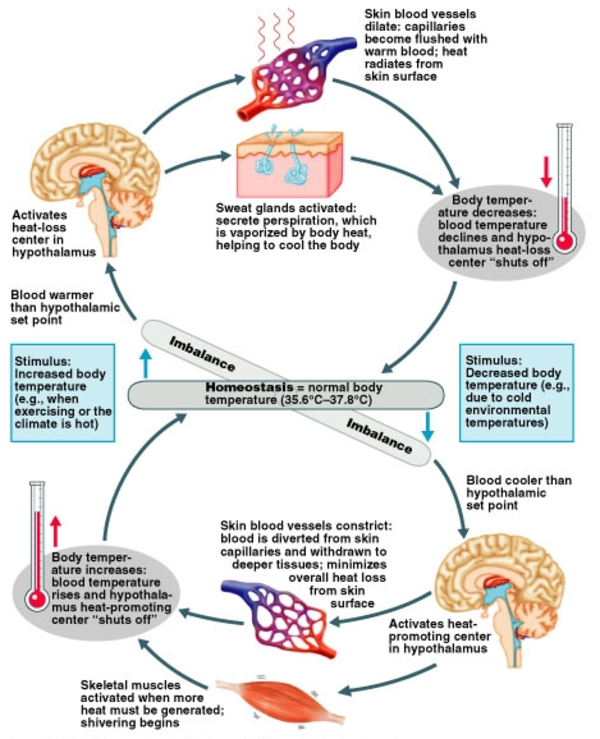
HUMAN BIOLOGY / THERMOREGULATION - Pathwayz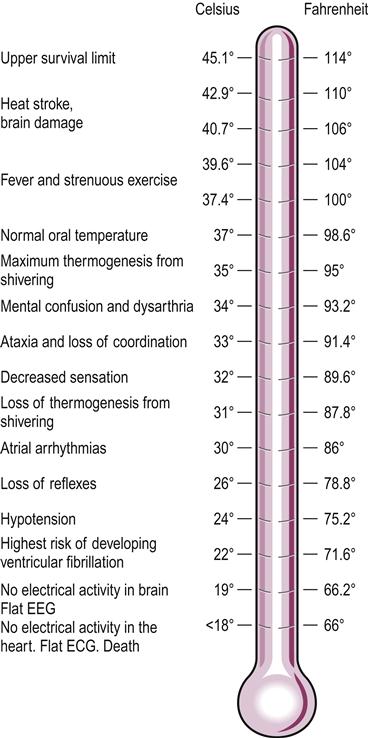
Thermoregulation: considerations for aging people | Musculoskeletal Key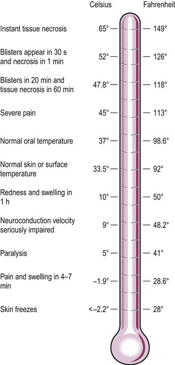
Thermoregulation: considerations for aging people | Musculoskeletal Key
Fundamental Concepts of Human Thermoregulation and Adaptation to Heat: A Review in the Context of Global Warming
Thermoregulation and Exercise: A Review – Ostéopathe Shawinigan | Antoine Del Bello Ostéopathe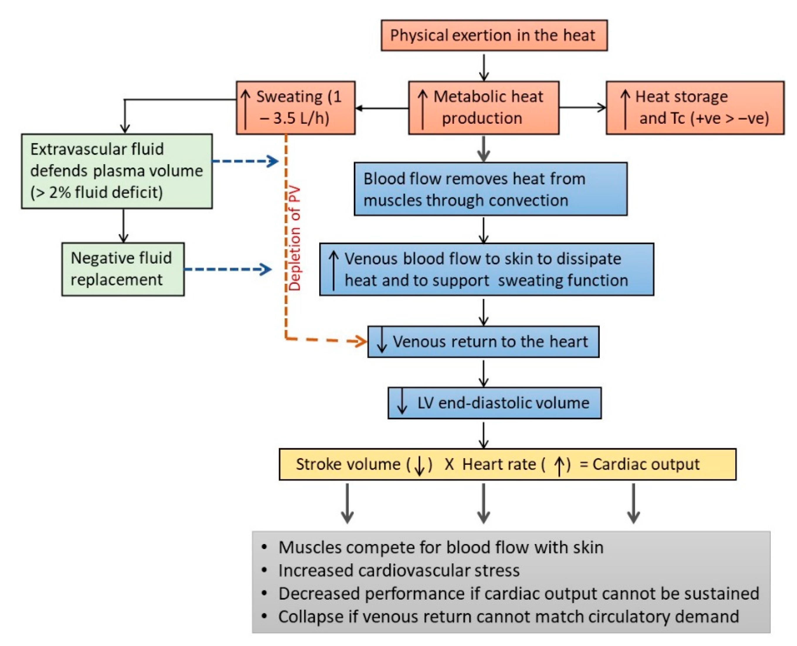
IJERPH | Free Full-Text | Fundamental Concepts of Human Thermoregulation and Adaptation to Heat: A Review in the Context of Global Warming
IJERPH | Free Full-Text | Limitations to Thermoregulation and Acclimatization Challenge Human Adaptation to Global Warming | HTML
Thermoregulation in humans - Wikipedia
The maggot, the ethologist and the forensic entomologist: Sociality and thermoregulation in necrophagous larvae - ScienceDirect
PDF) Limitations to Thermoregulation and Acclimatization Challenge Human Adaptation to Global Warming
Disposable low-cost cardboard incubator for thermoregulation of stable preterm infant – a randomized controlled non-inferiority trial - EClinicalMedicine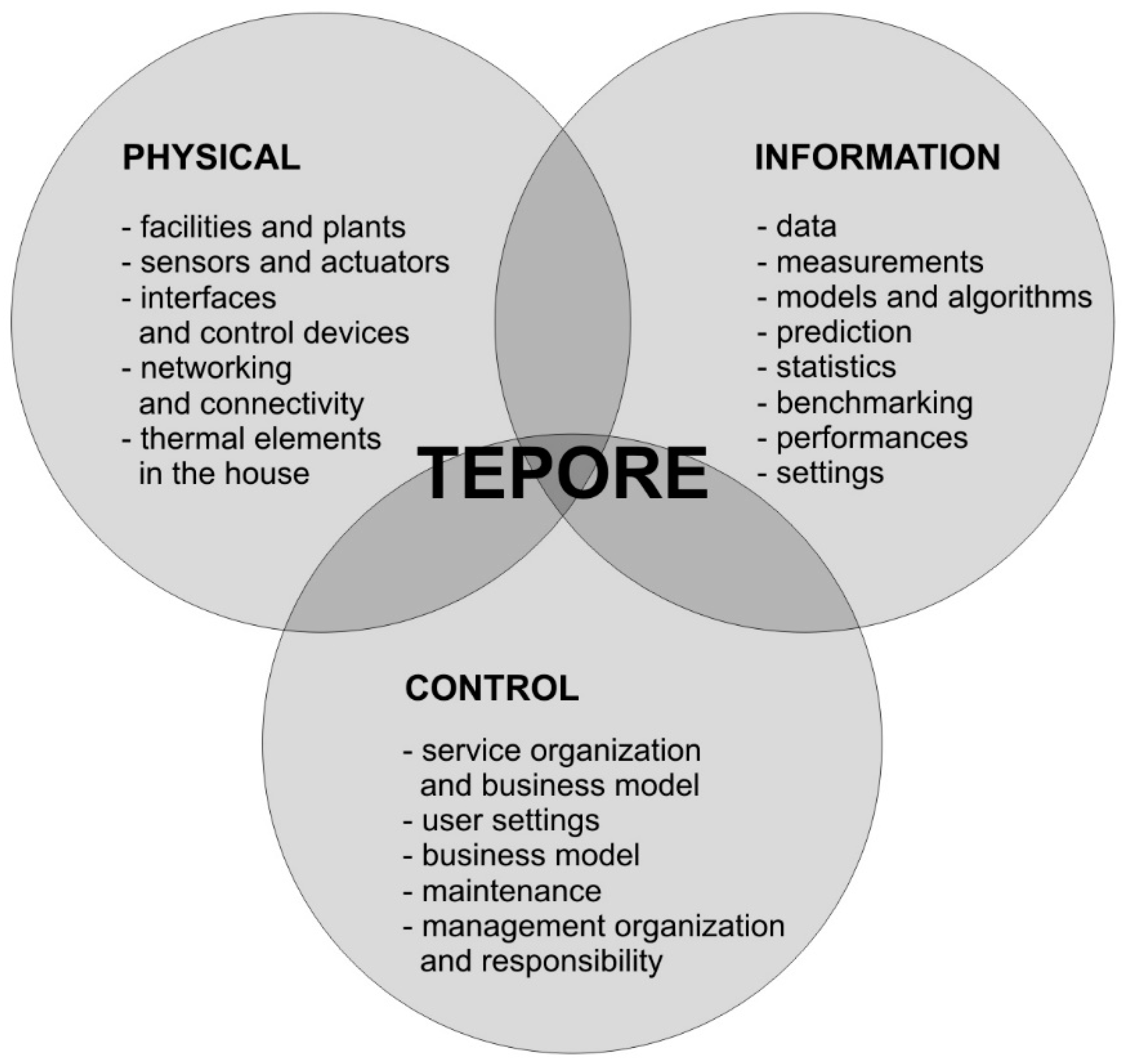
Sustainability | Free Full-Text | The Complexity of Simple Goals: Case Study of a User-Centred Thermoregulation System for Smart Living and Optimal Energy Use | HTML
PDF) Development of Thermoregulation Model of Surgical Patient and Heat Exchange with Air Condition in the Operating Room
Second graders and their teacher have PhUn learning about thermoregulation | Advances in Physiology Education
Improving Normothermia in Very Preterm Infants A Quality Improvement Toolkit
PDF) Thermoregulation, Energetics, and Behavior.
thermoregulation in animals class 12 | mechanism of thermoregulation in animals in hindi and urdu - YouTube
PERIPrem Bundle: Thermoregulation | West of England AHSN
The Thermoregulation Expert Map | Download Scientific Diagram
Fundamental Concepts of Human Thermoregulation and Adaptation to Heat: A Review in the Context of Global Warming
Temperature measurement and thermoregulation in the term and preterm infant
PDF) Limitations to Thermoregulation and Acclimatization Challenge Human Adaptation to Global Warming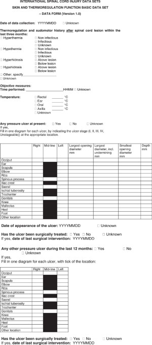
International Spinal Cord Injury Skin and Thermoregulation Function Basic Data Set | Spinal Cord
Vocal Function and Upper Airway Thermoregulation in Five Different Environmental Conditions | Journal of Speech, Language, and Hearing Research
STANDARD OPERATING PROCEDURES
Homeostatic disturbance of thermoregulatory functions in rats with chronic fatigue - ScienceDirect
Thermoregulation and Human Performance
Summary of a systematic review on maintaining normal body temperature (normothermia) - Global Guidelines for the Prevention of Surgical Site Infection - NCBI Bookshelf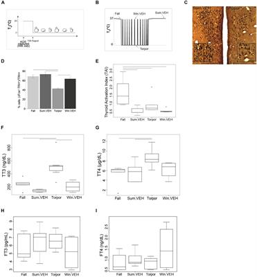
Frontiers | The Raphe Pallidus and the Hypothalamic-Pituitary-Thyroid Axis Gate Seasonal Changes in Thermoregulation in the Hibernating Arctic Ground Squirrel (Urocitellus parryii) | Physiology
PDF) Development and Validation of the Social Thermoregulation and Risk Avoidance Questionnaire (STRAQ-1)
Newborn Thermoregulation at birth Essay Example | Topics and Well Written Essays - 1750 words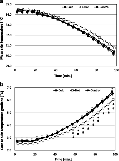
Nonthermal sensory input and altered human thermoregulation: effects of visual information depicting hot or cold environments | SpringerLink
Thermotaxis is a Robust Mechanism for Thermoregulation in Caenorhabditis elegans Nematodes | Journal of Neuroscience
Thermoregulation and Exercise: A Review – Ostéopathe Shawinigan | Antoine Del Bello Ostéopathe
Temperature regulation in women: Effects of the menstrual cycle: Temperature: Vol 7, No 3
thermoregulation | The brain is sooooo cool!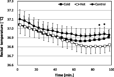
Nonthermal sensory input and altered human thermoregulation: effects of visual information depicting hot or cold environments | SpringerLink
Defects in Breathing and Thermoregulation in Mice with Near-Complete Absence of Central Serotonin Neurons | Journal of Neuroscience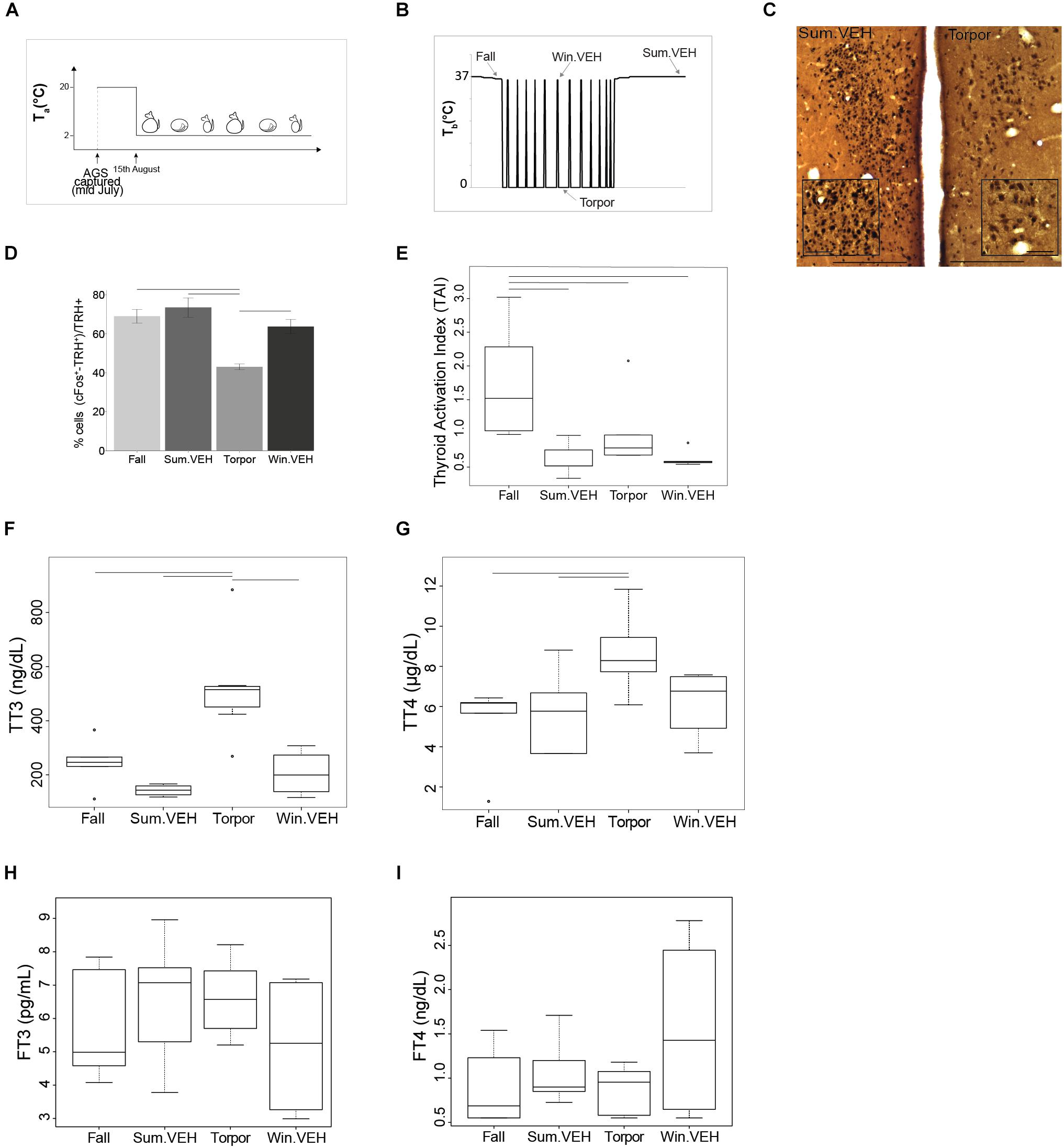
Frontiers | The Raphe Pallidus and the Hypothalamic-Pituitary-Thyroid Axis Gate Seasonal Changes in Thermoregulation in the Hibernating Arctic Ground Squirrel (Urocitellus parryii) | Physiology
 Thermoregulation: considerations for aging people | Musculoskeletal Key


Posting Komentar untuk "thermoregulation is the responsibility of:"